Study finds babies of anaemic mothers have smaller brains
A new study suggests that babies born to mothers with anaemia may have smaller brains, especially in areas involved in movement, learning and emotional control.
Scientists said the differences were visible by the time infants reached one year old and may contribute to difficulties with thinking and learning once children begin school.
Anaemia affects more than one in three pregnant women around the world. It is often caused by iron deficiency, which reduces the number of healthy red blood cells in the body. Common symptoms include tiredness, breathlessness and light-headedness. The condition is particularly widespread in sub-Saharan Africa and south Asia.
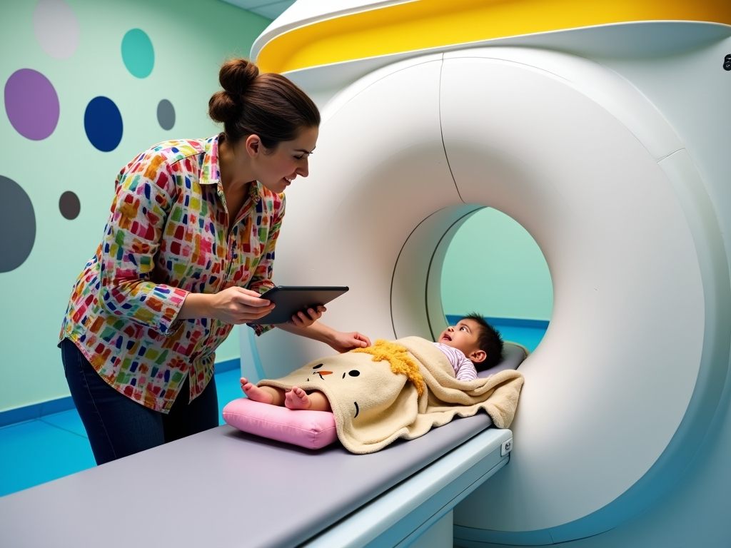
A team from King’s College London and the University of Cape Town monitored more than 300 mothers and their children in Cape Town. The babies underwent repeated brain scans from around three months of age until they were two years old.
On average, infants whose mothers had anaemia had total brain volumes about 4% smaller than those whose mothers did not have the condition. All of the mothers diagnosed with anaemia had mild cases.
The researchers identified differences in three particular brain structures: the putamen, the caudate nucleus and the corpus callosum.
Jessica Ringshaw, the study’s lead author and a researcher at King’s Institute of Psychiatry, Psychology & Neuroscience and the University of Cape Town, said these areas are involved in important neuropsychological abilities, including processing speed, emotional regulation and executive control.
The difference between the two groups appeared to grow during the first two years of life. For example, the corpus callosum was roughly 4% smaller at 12 months among babies born to mothers with anaemia, increasing to about 6% by 24 months.
Previous studies in South Africa involving toddlers and school-aged children have reported similar variations in brain size.
Ringshaw said the evidence indicated that the children were not simply catching up as they grew. The differences could still be present by the age of six, when children start school and may begin to experience clearer cognitive challenges.
She added that the results may help explain earlier research showing that children born to mothers with anaemia tended to perform less well on developmental assessments.
The mothers with anaemia and those without it were otherwise broadly alike in factors including household income and access to food, Ringshaw said. This made the researchers more confident that anaemia during pregnancy was linked to the brain differences they found.
The findings underline the need to prevent and treat anaemia among women of childbearing age, she said. Screening for anaemia and iron deficiency, especially during pregnancy, should be expanded, with oral or intravenous iron treatment used when needed.
Anaemia can have several causes, including infectious illnesses. The study found that anaemia was more common among women living with HIV, highlighting the importance of combining iron treatment with appropriate management of infections.
Reducing anaemia has been identified as a major global health goal, but progress has slowed over the past 20 years.
Ringshaw said anaemia must become a stronger public health priority to ensure that children have the chance not only to survive, but also to develop well and flourish.
The study, published in Brain Communications, used newer MRI devices designed to be less expensive and easier to transport than standard hospital scanners.
Researchers compared scans from conventional high-field MRI systems with images produced by portable low-field machines. They found that the smaller devices could identify the same brain differences in infants born to mothers with anaemia.
Steve Williams, a professor of neuroimaging at King’s, said the research showed that the newer technology could assess how malnutrition affects developing brains on a large scale, even in difficult settings. He said it could also support early and sensitive evaluation of health interventions.